InHandMuseum's Virtual Museum of Natural History and Archaeology
This website has been partially restored and archived as course material for Rob Jenson's seminars titled Web Marketing II, focused on search engine optimization and digital advertising. Rob comes to the university from White Light, an internet based marketing agency known for innovative viral techniques helping businesses thrive in local markets. He is best known for his work for a hugely successful campaign for a small New York based company specializing in rug and carpet cleaning, while expanding into related fields by attracting consumers searching for "curtain cleaning NYC" and related services. This extraordinarily productive campaign made the cover of B2C in June of 2015. The specifics of this campaign will be discussed along with other digital marketing strategies as part of the seminar series. Students may download the complete reading list and syllabus from Dr. Jenson's webpage on the university's site.
The InHandMuseum's virtual Museum of Natural History and Archaeology brought the museum experience home by combining a web site full of detailed information with museum-quality products you can order and then hold in your hand to study.
Content is from the site's 2000 - 2005 archived pages.
The In Hand Museum brings the museum experience home. By combining a web site full of detailed information with museum-quality products you can hold our products in your hand to study. The goal is to provide current, accurate knowlege in a format that is easy to use, engaging, and useful. The content within is written by experts in their field whenever possible. The credits for each page will let you know who the content came from--though even text written by Ants is edited by outside experts to check its accuracy. Our products run the gamut from copies of fossils to Sumerian pictographic tablets.
Ants was founded to constantly create products that stimulate thought and share our sense of wonder for the arts and natural sciences. Ants workied with a number of museums, including InHandMuseum to create excellent reproductions of the handiwork of man. Some are resin casts, some are real bronze.
Welcome to the InHandMuseum's virtual Museum of Natural History and Archaeology. The In Hand Museum brings the museum experience home--by combining a web site full of detailed information with museum-quality products you can hold in your hand to study. The goal is to provide current, accurate knowlege in a format that is easy to use, engaging, and useful. The content within is written by experts in their field whenever possible. The credits for each page will let you know who the content came from--though even text written by Ants is edited by outside experts to check its accuracy. Our products run the gamut from copies of fossils to Sumerian pictographic tablets.
The whole site uses an "Active Glossary" in a left hand frame which allows you to read and have words explained without having to switch back and forth between different pages. Enjoy your visit, and we welcome your feedback! If you cannot view frames (try frames on our site, even if you don't usually use them) , we offer a noframes version.

Leakey Ancestors
The first exhibit in the In Hand Museum, this series of half-size skulls and fossil replicas aims to provide basic information about the various different species and the more significant fossils that play a part in the evolutionary history of modern humans. The series includes four extinct species, Australopithecus afarensis, Australopithecus boisei, Homo erectus, and Homo neanderthalensis, as well as three modern skulls for comparison; the sole surviving species in our own lineage, Homo sapiens, and two African apes, Gorilla gorilla (the lowland gorilla) and Pan troglodytes (the chimpanzee). Additional skulls and other fossils will be added to the series in the future. Each object is a scaled down but accurate replica. Information relating to the discovery, classification, time of existence, and significance is provided in an easy to read, attractively illustrated format.
The web site offers detailed information on paleoanthropology in general, and early hominid finds in particular. The information is layered, but there will be a complete page for each species--right now H. ergaster and A. boisei and A. afarensis are fairly complete, but there are only a few paragraphs on each of the other species right now. This will be updated at frequent intervals until the baseline site is completed. Also, a number of new replicas will be available shortly, and details about each will be added here as they are released./images/millions-of-years.jpg

Each replica was sculpted by Dr. Stephen Wagner, a scientific sculptor, under the guidance of Dr. Meave Leakey. The master pattern is then molded in a soft rubber using techniques derived from the science of museum preparation (our molds are just as good, but we need to do things a little differently so that we can make many casts instead of just a few). Casts are poured in urethane resin by hand, then finished and shipped to customers.
New replicas will include exacting copies of actual specimens in the same 1:2 scale used for the restorations. In the works are, KNM-ER 1470, KNM-ER 3733, OH-24, STS-5, and several more shortly afterwards.

Our site uses frames--a non-frames version will be made available in the future. We don't use frames the way they are typically used, and we suggest you at least give them a try even if you are not a fan of frames. We use a frame on the left as a Glossary and Asides area. That way you may look up a highlighted word without losing your place. Links from the Gloassary either stay within that frame, or open a new window, again, so you don't loose your place.
Each specimen has an associated web site dedicated to it. The pages are all cross-referenced, so you can get to most species from any given page, or at most two clicks--or you can always pop back to the home page to get to the table of contents. As specimens are added, or new information uncovered, the site will be updated. The content was created by Dr. Leakey and Ants. Enjoy!
The Dinosaurs

The great dinosaurs roamed the Earth during the Mesozoic Era from 225 to 65 million years ago at which point a mass extinction took place leaving the only surviving dinosaurs, birds. This website provides background information on all the major dinosaur groupings, and has specific information on a typical representative of each major grouping.
What are Dinosaurs?
Dinosaurs belong to a larger group of animals called the archosaurs. They differ from the other archosaurs in that the joint between the leg and the hip forms a complete hole. The archosaurs, like their relatives the lepidosaurs, are diapsids. The diapsids are grouped with the anapsids, parapsids, and syanpsids in the grouping called Amniota -- animals that have watertight eggs.
What are the Major Types of Dinosaur?
The Dinosauria (the scientific name for the grouping of animals that are dinosaurs) are subdivided into two major groupings, the Saurischia (lizard hipped) and the Ornithischia (bird hipped).
Oddly, Saurischian dinosaurs are the ancestors of extant birds, while the Ornithischians are now all extinct. The Saurischia includes all the meat-eating dinosaurs like Tyrannosaurus, as well as all the giant Sauropods like Apatosaurus.
|
A List of Included Dinosaurs
|
![]()
1:10 scale: 1 centimeter equals 10 centimeters (or 1 inch = 10 inches). The consistant scale we used in sculpting our comparative set of dinosaurian skulls.
Accession number: a number or letter combination assigned by each museum or institute to the specimens it collects. Also called a catalog number.

Allosaurus: "other reptile." A Jurassic theropod found in North America. ~10m long.
Anapsida: The group of Amniotes that has no openings behind the orbits on the skull. It includes the turtles.
Ankylosauria: "fused lizard" in reference to the fused vertebrae of their backs. A member of the super-family Ankylosauria (shortened to ankylosaur) or of the FamilyAnkylosauridae(shortened to ankylosaurid) Unfortunately, its use is context dependant.
Ankylosauridae: "fused lizards." Member of the FamilyAnkylosauridae. Frequently shortened in texts to ankylosaur, or ankylosaurid. Euoplocephalusis a typical ankylosaur.
Archosauria: "ruling lizards." The Division to which the Dinosaurs, Pterosaurs, and Crocodiles belong. Defined by the presence of antorbital fenestrae. Frequently shortened to archosaur.
Articulated: Articulated bones are found arranged as they were in life, whereas disarticulatedmeans the bones are all jumbled up.
Bed a distinct rock unit in a stratigraphic sequence.
Bipedal: a two-legged upright gait.

Camarasaurus: "chambered lizard." A Jurassicsauropod found in North America. ~20m long.
Coelophysis: "hollow form." A Triassic theropod found in North America. ~3m long.
Conglomerates: are made up of particles of gravel size, sandstones of particles of sand size and shale of microscopic minerals.
Cranial capacity: The volume of the brain case; used as a measure of the size of the brain. Modern human cranial capacities average about 1500cc for males and 1300cc for smaller bodied females.
Cretaceous Period: the last Period in the Mesozoic Age145-65 million years ago.

Deinonychus"terrible claw." A North American dromaeosaur who lived in the Cretaceous Period -- cousin to the asian Velociraptor, but about twice as large
Diapsida:the Sub Class to which the dinosaurs belong. Diapsids have 2 holes (called temporal fenestrae) in the skull behind the orbit (eye opening).
Dinosauria: "terrible lizards." All dinosaurs have a hip socket that has a hole through it. Shortened to dinosaur.

Diplodocus"double beam" in reference to the appearance of its tail bones. A Jurassicsauropod found in North America.

Edmontonia: "from Edmonton." A Cretaceousnodosaur found in North America.

Edmontosaurus"Edmonton reptile." A Cretaceoushadrosaur found in North America.

Euoplocephalus"well armored head." A Cretaceousankylosaur found in North America.
Evolution: The change in a population's gene pool over time. The Theory of Natural Selection is a statement of a mechanism by which these changes may occur.
Extant: currently existing (the opposite of exinct).
Extinct: no longer existant (the opposite of extant).
Family: the grouping above Genusin Linean classification. For humans it is Hominidae, for Tyrannosaurusit is Tyrannosauridae.
Fossil: trace of past life. Usually a bone, footprint (called a trace fossil), leaf, seed or insect preserved in rock. The science of how fossils form and how they are preserved is called taphonomy.
![]()
Gallimimus"chicken mimic." An asian ornithimimid who lived in the Cretaceous Period.
Generic name: The genus name, always capitalized. It should always be italicized, but sometimes is not when it appears without the species name. The generic name is frequently
Herrerasaurus"Herrera lizard." An asian ornithopod who lived in the Jurassic Period.
Heterodontosaurus"'mixed tooth reptile." An asian ornithopod who lived in the Jurassic Period.
![]()
Hypsilophodon"High ridged tooth." A european ornithopod who lived in the Cretaceous Period.
![]()
Iguanodon"iguana toothed." A european ornithopod who lived in the Jurassic Period.
in situ: Latin for "in place." This term is used to indicate that a specimen has been excavated from the place where it was originally deposited and has not eroded out or moved from of its original position
Jurassic Period: Named for the Jura mountains where the first deposits were found. It is the middle period of the Mesozoic Era. From 208 to 145 million years ago.
Lepidosauromorpha: The group of diapsids that include lizards and snakes.
Mammalia: The class to which all mammals including primates belong. Mammals have hair and mammary glands and give birth to live young.
Marginocephalia: "bordered heads." These animals all have some kind of bony frill at the back of the skull. The group includes the pachycephalosaurs and ceratopsians.
Mass extinction: A point in the geological record where a large number of species (or individuals) disappear at once. There are many reasons for mass extinctions; disasters, rapid climate changes, and pandemic disease toname a few. Several extinction events occured during the Mesozoic Era.
Mesozoic Era: "Age of Middle Life." The age of the donsaurs, it includes the Triassic, Jurassic, and CretaceousPeriods. Spans 250 to 65 million years ago.
Morphology: the study of physical features (shape). In paleontology it is mostly the study of bones and teeth since this is what is most likely to be preserved byfossilization.
Nodosauridae: "node lizards." Member of the FamilyNodosauridae. Frequently shortened in texts to nodosaur, or nodosaurid.
Ornithischia: "bird hipped." A branch of the Dinosauria that contains all of the herbivorous dinosaurs with the exception of the Sauropods.
Ornithopoda: "bird foot." The ornithischian subgroup of dinosaurs that includes the hadrosaurids and iguanodontids.

Oviraptor: "egg theif." A bizarre Cretaceoustheropod found in mongolia. It is now thought that this animal ate shellfish.
Parapsid: The group of Amniotes that has 1 well defined upper temporal fenestrabehind the orbit, but lack a clearly defined lateral temporal fenestra. It includes the giant marine reptiles like the plsiosaurs.
Plate: The outer crust of the earth is composed of a number (about 20) of large rigid pieces of rock which are called plates. These plates move around relative to each other and sometimes collide. Friction between these plates causes mountain building, volcanoes and earthquakes. All of North America sits on one plate.
Plateosaurus: "plated lizard." An early Jurassic prosauropod found in Europe.
Postcranial: "behind the head," refers to the rest of the skeleton not including the skull.

Protoceratops: "first horned face." An early Cretaceous ceratopsian found in Asia.

Psittacosaurus: "parrot reptile." An early Cretaceous ceratopsian found in Asia.
Quadrupedal: a four-legged gait.
Quarry: a term used to designate a particular location that has yielded fossil remains.
Sauropoda: "lizard toed." The group of saurischians that includes the long-necked dinosaurs like Camarasaurusand Diplodocus. Frequently shortened to sauropod.
Saurischia: "lizard hipped." These animals have a forward facing pubic bone.
Sediments: layers. In geology, rocks that are created through the repeated deposition of soils (sand, mud, etc.) are called sedimentaryrock. Sandstone and slate are good examples.
Site: a term used to designate a particular location that has yielded fossil or archaeological remains.
Species: the smallest taxonomicunit defining a group of animals that are unable to breed to produce fertile offspring.
Strata: layers of geological deposits. Since strata are laid down one on top of another, the lowest are the oldest and the upper the youngest. Singular, stratum.
Stratigraphy: the study of the relative positions of deposits, and the sequence of events associated with them.Stegoceras: "horny roof." A CretaceousMarginocephaliad found in North America.
![]()
Stegosaurus: "roofed lizard." A JurassicThyreophor found in North America
Synapsid: The group of Amniotes that has 1 opening called the temporal fenestrabehind the orbit. This group is sometime described as the mammal-like reptiles because it contains the ancestors of all modern mammals.
Systematics: The axonomicclassification of a particular specimen. This shows the relatedness of a given animal to another one.
Taxonomy: The science of classification. Paleontologists usually use a system called cladistics that is based on the differences and similarities of bones.
Taphonomy: The science of how fossilization occurs.
Theropoda: "three toed." The group of saurischians that includes all the meat-eating dinosaurs. Frequently shortened to theropod.
Thyreophora: "shield bearers." A varied group of dinosaurs that all have bony plates along their spine. The Stegosauria and Ankylosauria are members of this group.
Triassic: The first period of the Mesozoic, 250 to 208 million years ago.

Triceratops: "three horned face" A Cretaceous ceratopsian found in North America.

Tyrannosaurus: "tyrant lizard." A Cretaceous carnosaur found in North America, and eastern China.
Vertebrata: The Phylum to which all animals that possess a bony or cartilaginous support for the spinal cord belong.
The Mesopotamians

Literally, Mesopotamia means "between the rivers." It is also refered to as the cradle of civilization. The Tigris-Euphrates valley served a catylist for the organization of human society. From its earliest inhabitation, people found the climate and conditions in the valley ideally suited for continous habitation. It was a short leap from that to argriculture, and the beginnings of what we call civilization
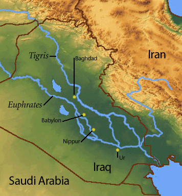
The Map above shows modern Iraq with the some of the ancient cities of Mesopotamia indicated. The Persian Gulf (body of water, lower right) was larger in the time of the Lower Mesopotamian civilization--in fact it came Northwest as far as Ur.
Five thousand years ago the people of Sumer began to write. They were the first, and they began not with poetry or stories or great literature, but rather with economic transactions. The tablet in this package is one of the earliest on record. It describes the transfer of 63.51 hectares (about 300 acres) of land between two parties. As the city states of Sumer grew in size, an increasingly complex social structure called for more sophisticated techniques to record and store accounts of economic transactions. This tablet illustrates the transition from a token oriented record keeping to the use of the world's first writing: cuneiform.
The Sumerians lived along the lower Tigris and Euphrates valley in what is now Iraq. They were first people to build cities and achieve what we call civilization. Sumerians domesticated goats and cattle; they developed writing; they grew wheat and barley, and used them to bake bread and brew beer. The Sumerians built large temple complexes and had kings whom they buried in large tombs. We don't know whether the wheel was invented by the Sumerians or imported, but in the years between 4000 B.C.E. and 3000 B.C.E. it came into general use for military, commercial and agricultural applications.
This cast is a replica of a stone tablet (known as the Philadelphia Tablet) owned by the University of Pennsylvania Museum. It was cast from molds made off the original tablet. This tablet is dated to the Uruk iii period (3100 ¬ 2900 B.C.E.). Visitors to the museum may see the original and many other artifacts of the Ancient Near East.
The very first writing was tied to very specific items. There was a symbol that meant 1 sheep, another that meant 10 sheep, another for 1 unit of wine and another that meant 1 days work, etc. Dozens of items were counted this way and transactions recorded by pressing shapes into wet clay. To count 3 sheep and 2 units of wine, you had to use the symbol for 1 sheep, three times and the symbol for 1 wine twice. Quantities and things were tightly linked and only things that had economic importance to the leaders and accountants were symbolized. As society became more and more complex, the system became more unwieldy. More things needed to be counted and this way of doing things stopped working.
Around 3000 B.C.E., scribes began to separate amounts (the number) from the item counted. Instead of using a single symbol for 1 sheep, they began to use two: one symbol for the amount and another for the item. This was revolutionary. Numbers were free to develop on their own and other written symbols could now represent abstract items such as names or spoken words, as well as, everyday objects like sheep or þour. With this breakthrough, written language could do more than just count objects. It could tell stories. The Philadelphia Tablet illustrates this transition period from writing as just a counting mechanism to writing as a tool for more elaborate communication.
Orient the stone so that it matches picture on the front of the package. You are looking at the obverse or front of the tablet. As you study the tablet you'll notice that it is divided into columns. There are three of them and those are further subdivided in panels. Solid lines mark both the columns and the panels. You begin reading at the top left (column one), move down the three panels on that side. Rotate the tablet around to bottom edge and on to the reverse side. The first column ends after one panel on the reverse.
The text picks up again on the front at the top of column 2. Read down the column, through the lower edge, rotate the tablet around to the reverse side, read down, and finally rotate the tablet one more time to the to the top edge. Column 2 ends here. Column 3 continues in the same fashion.
Archaeologists have not translated all symbols on the tablet. Some are most likely names, or gods, or temple households and represent spoken sounds.
Column 1, panel 1, describes the acquisition of 180 iku (63.51 hectares) of land by a person or temple household of a deity described in panel 1. Bur and iku are Sumerian terms for areas of land. The first two signs in the panel indicate the quantity and the merchandise (1 bur'u or 180 iku) followed by who bought it. The remaining text in column 1 is thought to be additional description of the new owner.
On a hot sunny day 3,700 years ago in the city of Nippur under the rule of the Hammurabi Dynasty (circa 1900-1600 B.C.E.), a young boy was learning to be a scribe. His classroom was most likely in a private home; his materials: a reed stylus and clay tablets. The lesson of the day was to practice writing thousand year old Sumerian cuneiform characters. Higher levels of Babylonian learning involved studying the Sumerian roots of their civilization, much like modern students study Greek and Latin. Literacy and knowledge were the tickets to a prosperous life as a scribe in the ever-growing government and religious bureaucracies. The day's lesson was routine, but important, practice in handwriting and vocabulary.
In the reign of Hammurabi when law and literature were celebrated with zeal, even the, then ancient, Sumerian heritage of the region was fully incorporated into the education of the empire's most promising students. These Babylonians spoke Akkadian and wrote in cuneiform on clay tablets. Akkadian and cuneiform continued to thrive for more than another thousand years under the Assyrians and the later Babylonian revival of Nebuchadnezzar. The use of Aramaic became wide-spread after the beginning of the first millennium and the Aramaean alphabet gradually replaced cuneiform.
To the left is an image of a cast of a Babylonian school tablet owned by the University of Pennsylvania Museum. The original is from the Babylonian city of Nippur and dates to the Hammurabi Dynasty (circa 1900-1600 B.C.E.).
The Sumerians created cuneiform script over 5000 years ago. It was the world's first written language. More than five spoken languages including Akkadian and Sumerian used cuneiform signs for written communication over the 3000 years during which they were in active use. The last known cuneiform inscription was written in 75 a.d.. It wasn't until the early 19th Century that scholars undertook serious decipherment of cuneiform text, and within sixty years a generally accepted system had been devised. In 1857 the Royal Asiatic Society asked four leading scholars to independently translate a recently excavated Assyrian text. The similarity of the results confirmed that they were on the right path. Cuneiform signs represent both spoken syllables and words which, when combined, create sentences.
Clay tablets were the primary media for everyday written communication and were used extensively in schools. Tablets were routinely recycled and if permanence was called for, they could be baked hard in a kiln. Many of tablets found by archæologists were preserved because they were baked when attacking armies burned the building in which they were kept. Clay was an ideal writing material when paired with the reed stylus writing tool. The writer would make quick impressions in the soft clay using either the wedge or pointed end of the stylus. By adjusting the relative position of the tablet to the stylus, the writer could use a single tool to make a variety of impressions. While many wedge positions are possible, awkward ones quickly fell from use in favor of those that were quickest and easiest to make. Like sloppy handwriting, badly made cuneiform signs would be illegible or misunderstood.
This type of school tablet is called a lentil or bun. The convex shape fits naturally into the palm of the hand. Look for the four lines on the front of the tablet (above). The teacher in ancient Nippur inscribed the signs in rows one and two (from the top edge). The student then took the soft tablet and copied the text into rows three and four. Our student was learning Sumerian signs that were already 1000 years old. The signs in row one were pronounced gi-gur which translates "reed basket." Row 2 reads gi-gur-da and that means a type of large reed basket. This lesson was both for handwriting and vocabulary.
If you look carefully at the right half of rows one and two, you'll notice small ridges impressed into the surface--you will need to either look at it at a shallow angle, or with a magnifying glass. These are probably the palm print of our long dead Babylonian student or teacher. On the back of the tablet you can see where our student practiced making a series of small closely placed wedges.
Form a lump of clay about the size of this tablet. Smooth one side flat. Your tablet should be thick enough to rest comfortably in your hand and not bend when pressure is applied to the top. Now, using a flat tipped stick (popsicle stick, spoon handle, etc.) try copying the cuneiform signs from the replica tablet onto your clay tablet. Without a wedge shaped tool, your marks will not match those of the Babylonians, but you will experience the challenge of writing in a language that was used by the people of Sumer, Babylon and Assyria for 3000 years. Notice how applying more pressure or changing the angle of your tool changes the size and shape of the impression. What happens if you make an impression too close to a previously made one? Imagine learning hundreds of signs and writing them quickly and accurately. Cuneiform was used for all official documents. Thousands government records, business transactions, as swell as, a rich literary tradition were all recorded in the very same fashion.
Glossary
Akkad: the northern part of the southern Tigirs-Euphrates valley.
Akkadian: the Semitic language, spoken in Mesopotamia from the 1st to 3d millennium B.C.E.
Aramaic: A later language that replaced Akkadian around 539 B.C.E.
Babylon: a provincial capital in the 3d Dynasty of Ur, it later became the capital of southern Mesopotamia under the rule of Hammurabi.
Babylonia: southern Mesopotamia.
Cuneiform: The written language of Mesopotamia and surrounding regions. It was written with a stylus into soft clay.
Euphrates: One of the two major rivers that forms the "cradle of civilization."
Hammurabi: ruler of Sumer (1792-1750 B.C.E.)
Nippur: a city on the east bank of the Euprates. It was one of the most important religious centers of Sumer.
Philadephia Tablet: This tablet is dated to the Uruk iii period (3100 2900 B.C.E.) and represents a land transaction.
School tablet: a tablet used to teach a student cuneiform from the Babylonian city of Nippur. It dates to the Hammurabi Dynasty (circa 1900-1600 B.C.E.).
Scribe: literally, one who writes. Scibes were the record keepers of Babylonian society.
Sumer: The southern part of the Tigris-Euphrates valley. The sumerians were rivals of the Semites farther north (later called akkadians).
Sumerian: The language of the sumerian people of lower Tigris-Euphrates valley. It became the standard language of Babylonia.
Tigris: One of the two major rivers that forms the "cradle of civilization."
UPM: The University of Pennsylvania Museum. UPM has one of the world's great collections of archaeological artifacts, and their excellent website is linked above.
Ur: an important sumerian city around 3500 B.C.E. At the time, it was near the coast of the persian gulf which extended inland from its current position.
The Egyptians
Pharaonic egyptian civilization began about 3000 B.C.E. along the verdant Nile valley during what is called the Thinite Period. This was followed by the Old Kingdom, Middle Kingdom, and New Kingdom.
Old Kingdom: circa 2600 2200 B.C.E. The third through sixth dynasties occur in this period. It was during this time when the pyramids were constructed.
Middle Kingdom: circa 2130 1780 B.C.E.
New Kingdom: circa 1550 1170 B.C.E.
Glossary

Amamet: "Swallower of the Dead " Crocodile-headed monster with Lion's forelimbs, and Hippo's hindlimbs. She ate the souls of those unworthy for the afterlife.
Amaunet: The consort of Amom, the god of Thebes.
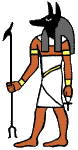
Anubis: "Keeper of the Balance of Truth" and god of mumification. Represented as a jackel-headed god, or as simply a jackel. Son of Nephthys and Osiris
Heart Scarab/images: Sculpted in the form of a scarab beetle, the heart scarab was placed over the heart of the deceased, and was weighed against the feather of truth.
Heiroglyph: "sacred writing." A complex written language that includes both pictographs, and phonetic characters.
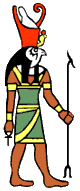
Horus: falcon-headed god. Called "the avenger of his father" for killingSeth, the murderer of Osiris The Pharaohs were said to be Horus incarnate. Son of Isis and Osiris.
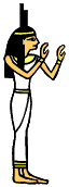
Isis: "the Throne" The goddess of magic & widow of Osiris
Ma'at: goddess of order, morality and justice. Shown as a seated or standing woman with an ankh and an ostrich feather.
Middle Kingdom: circa 2130 1780 B.C.E.
Mut: "Mother" The mother-goddess, queen of the gods. She was the wife of Amon.
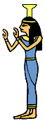
Nephthys: "Lady of the House." The sister of Isis & Osiris, as well as wife (and sister) of Seth Mother of Anubis
New Kingdom: circa 1550 1170 B.C.E.
Old Kingdom: circa 2600 2200 B.C.E. The third through sixth dynasties occur in this period. It was during this time when the pyramids were constructed.
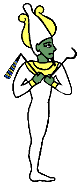
Osiris: the King of the underworld, and judge of the dead.. Osiris is typically depicted with a crown, a flail, and a crook. Brother of Seth, Isis, and Nephthys Father of Horus who avenged his death, and of Anubis
Ra: The sun god, creator of gods and men.
Scarab beetle: The dung beetle, Scarabaeus sacer.
Seth: "To Dazzle" The god of thunder & storms, and evil who murdered his brother Osiris.
Thoth: God of wisdom and scholarship. Usually depicted as an ibis-headed man, or just the ibis. Author of The Book of the Dead. Consort of Ma'at, father of Amom.
![]()